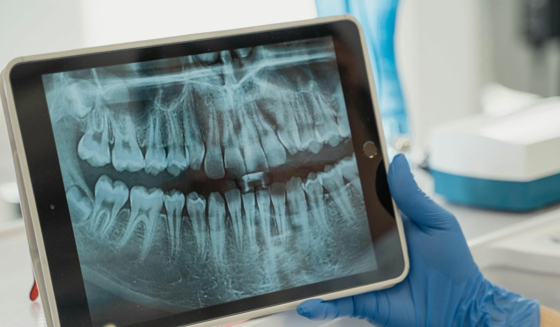
The discussion around routine dental X-rays often centers on the frequency of exposure or the cost, yet it frequently misses the central point: the indispensable nature of these images for a thorough assessment of oral health. The notion that a visual examination alone is sufficient for comprehensive dental care is deeply flawed; a mirror and a probe simply cannot penetrate the cortical bone and enamel to reveal the true state of the dental structures. To rely solely on a direct view is to ignore nearly half of the potential pathology brewing within the oral cavity. What the human eye registers is merely the surface—the crown of the tooth and the visible gum tissue—a superficial landscape that belies the complex, hidden activity underneath. This underlying complexity includes caries beginning in the tight spaces between teeth, the subtle erosion of the alveolar bone that signals aggressive periodontal disease, and the slow, silent growth of cysts or other pathologies within the jawbone. Dental radiography, therefore, is not an optional add-on but a fundamental extension of the dentist’s ability to see and diagnose. Without this crucial layer of visual data, the clinician is operating with an incomplete map, significantly increasing the likelihood of delayed diagnosis and more invasive, costly treatment down the road. The true value of an X-ray lies not just in what it finds, but in what it pre-empts.
A Mirror and a Probe Simply Cannot Penetrate the Cortical Bone and Enamel
The majority of clinicians use visual examination together with the dental explorer to decide if an occlusal surface is in need of restoration or if preventive management is required.
The limitations of a direct, visual-tactile examination become apparent the moment one considers the anatomy of the tooth. The majority of clinicians use visual examination together with the dental explorer to decide if an occlusal surface is in need of restoration or if preventive management is required; however, this traditional approach is prone to error, particularly when assessing the tightly packed interproximal surfaces where early decay often initiates. When two teeth abut closely, the light and instrument cannot effectively reach or reveal the incipient breakdown of the enamel and dentin. Furthermore, the presence of existing restorations (fillings, crowns) can mask underlying issues, making it impossible to detect recurrent caries or marginal breakdown simply by looking. Studies comparing visual findings with radiographic and even histological evidence frequently show a significant margin of error for visual inspection alone. This suggests that relying solely on the naked eye inevitably leads to missed diagnoses in the early stages, pushing the eventual treatment toward more extensive and complicated procedures, such as root canals or extractions, which could have been avoided entirely.
Where Early Decay Often Initiates
Bitewing X-rays: These X-rays detect cavities between teeth that may not be visible during a visual exam.
The Bitewing X-ray is the workhorse of preventative dentistry, specifically designed to circumvent the limitations of the visual exam. Bitewing X-rays: These X-rays detect cavities between teeth that may not be visible during a visual exam. By positioning the film or digital sensor parallel to the teeth and instructing the patient to bite down gently, this type of image provides a crystal-clear, cross-sectional view of the crowns of the upper and lower back teeth. It is precisely in the interproximal spaces, where food particles and plaque are most likely to linger undisturbed, that demineralization and carious lesions begin their silent progression. An X-ray image captures the change in radiodensity—the dark shadow of decay—long before it breaches the outer surface of the tooth and becomes clinically visible or palpable with a probe. This early detection capability is perhaps the single most compelling argument for their routine use, enabling the dentist to intervene with a small, conservative filling or even non-invasive measures like fluoride varnish, thereby preserving a maximal amount of natural tooth structure.
The Subtle Erosion of the Alveolar Bone
X-rays help detect signs of bone loss around the teeth, a key indicator of advanced periodontal disease.
Beyond simple cavities, radiography is the definitive tool for assessing the condition of the supporting structures, primarily the periodontium. X-rays help detect signs of bone loss around the teeth, a key indicator of advanced periodontal disease. Periodontitis—often referred to as gum disease in its more advanced state—is a chronic inflammatory condition that leads to the gradual destruction of the bone that anchors the teeth in the jaw. In its early stages, the clinical signs may be as mild as slight gingival inflammation or occasional bleeding, symptoms which are easy for a patient to dismiss. The damage, however, is occurring silently below the gum line, where it is completely invisible to a visual exam. The Bitewing and Periapical X-rays provide the vertical height of the alveolar crest and reveal the characteristic horizontal or vertical bone loss patterns indicative of the disease’s progression. Without this objective, radiographic evidence, a dentist is merely guessing at the extent and severity of the disease process, fundamentally hindering the ability to devise a proper periodontal treatment plan, which may range from scaling and root planing to surgical intervention.
The Condition of the Jawbone
X-rays also help assess the condition of the jawbone, including bone density, bone loss, and potential issues with the temporomandibular joint (TMJ).
The utility of X-rays extends well beyond the tooth itself to provide an essential panoramic view of the entire bony architecture of the face and mouth. X-rays also help assess the condition of the jawbone, including bone density, bone loss, and potential issues with the temporomandibular joint (TMJ). Larger films, such as a Panoramic X-ray (Panorex), are vital for surveying the general state of the mandible and maxilla, which is critical for identifying non-odontogenic pathologies. These images can reveal impacted wisdom teeth that are positioning themselves poorly and threatening adjacent healthy roots, or they can highlight cysts, tumors, or other bony lesions that have not yet caused external symptoms. Furthermore, the X-ray is an indispensable diagnostic aid for endodontic (root canal) and oral surgery procedures, providing the spatial map required to safely navigate the root anatomy and plan the precise placement of dental implants. It is a fundamental pre-requisite for any complex restorative or surgical intervention.
The Spread of Bacteria to Other Parts of the Body
An untreated dental infection can lead to serious complications, including tooth loss and the spread of bacteria to other parts of the body.
One of the most concerning and often unseen issues that X-rays address is the presence of periapical infections. An untreated dental infection can lead to serious complications, including tooth loss and the spread of bacteria to other parts of the body. These infections, which typically manifest as abscesses at the apex of the tooth’s root, are often the result of deep decay or trauma that has compromised the pulp (nerve and blood vessels) of the tooth. A visual exam may reveal nothing more than a discolored tooth or a subtle swelling, but a Periapical X-ray will clearly show the radiolucent (dark) area around the root tip—evidence of bone breakdown caused by the bacterial load. If left unchecked, these infections can spread locally through the jawbone or, far more seriously, lead to systemic health problems, a risk that underscores the importance of the X-ray as a tool for overall health screening, not just dental maintenance.
A Critical Part of the Preventative Health Strategy
Regular X-rays allow dentists to track the progression of dental conditions, such as tooth decay, gum disease, or bone loss, and assess the effectiveness of ongoing treatments.
The role of radiography is not confined solely to initial diagnosis; it is also a dynamic component of long-term patient care. Regular X-rays allow dentists to track the progression of dental conditions, such as tooth decay, gum disease, or bone loss, and assess the effectiveness of ongoing treatments. For patients with known periodontal disease or those undergoing complex procedures like orthodontics, serial X-rays provide an essential baseline and a means of objective comparison over time. This longitudinal data allows the clinician to monitor the stability of bone levels, assess the integrity of existing restorations, and detect any new or recurring areas of concern before they become symptomatic. This is a critical part of the preventative health strategy, ensuring that treatment is adjusted proactively rather than reactively, maximizing the longevity of the patient’s dentition.
Safety Measures
Dental X-rays are safe and are performed using low levels of radiation.
The concern over radiation exposure is a valid one, and one that is frequently addressed by advancements in dental technology. Dental X-rays are safe and are performed using low levels of radiation. Modern dental practices overwhelmingly utilize digital radiography, which requires significantly less radiation exposure—up to 80% less than older film-based X-rays—to produce a high-quality image. The radiation dose from a full set of modern dental X-rays is often comparable to the natural background radiation an individual receives simply by spending a few hours outside or flying on a short airplane trip. Furthermore, the decision to take X-rays is always based on the individual patient’s clinical needs, factoring in their history, age, and the visible findings, ensuring that the exposure is always as low as reasonably achievable, a principle known as ALARA.
Determining the Need for Surgery
X-ray helps to locate which teeth should be operated in order to restore lost bone tissues.
In the sphere of complex treatment planning, X-rays transition from a diagnostic aid to an essential surgical blueprint. X-ray helps to locate which teeth should be operated in order to restore lost bone tissues or to facilitate procedures like apicoectomies or regenerative periodontal surgery. For cases involving implant dentistry, advanced 3D imaging, such as Cone-Beam Computed Tomography (CBCT), which is a specialized form of X-ray, provides a detailed, three-dimensional view of the bone volume, nerve pathways, and anatomical landmarks. This high-resolution map is non-negotiable for safe and effective implant placement, drastically reducing the risk of complications and ensuring the long-term success of the prosthesis. The surgical plan moves from an educated guess to a meticulously planned procedure based on hard, objective radiographic data.
The Patient’s Understanding of Their Dental Condition
Providers are able to use visual output generated by AI and machine learning to better help patients visualize and understand their dental conditions.
Beyond the technical necessity for the clinician, X-rays play a vital role in patient education and communication. Providers are able to use visual output generated by AI and machine learning to better help patients visualize and understand their dental conditions because an X-ray provides undeniable, visual proof of an issue that the patient cannot otherwise feel or see. Showing a patient a large, dark shadow of interproximal decay or a clear image of their bone loss makes the abstract concept of ‘gum disease’ concrete and urgent. This visual evidence dramatically improves patient compliance with the recommended treatment plan, turning a potentially vague, abstract concept into a clear, shared reality. When a patient can see the hidden problem for themselves, they become a more invested and proactive partner in their own oral health journey, moving past the simple transaction of a check-up to a deeper understanding of their own body.
A Fundamental Extension of the Practitioner’s Eyes
The X-ray should be seen not as an ancillary task, but as a fundamental extension of the practitioner’s eyes, reaching into the anatomical spaces that light and mirror cannot touch.
Ultimately, the argument for the importance of dental X-rays is an argument for comprehensive care and early intervention. The X-ray should be seen not as an ancillary task, but as a fundamental extension of the practitioner’s eyes, reaching into the anatomical spaces that light and mirror cannot touch. It is the primary means of detecting early disease, quantifying the extent of deep pathology, and safely planning complex surgical procedures. By providing this indispensable subsurface visibility, X-rays move dental practice from reactive symptom management to proactive, preventative medicine, securing the patient’s long-term oral health and often preventing small issues from spiraling into significant pain and financial burden.
