Tooth decay doesn’t begin with pain. It starts as mineral loss on enamel surfaces. Acidic environments strip calcium. Saliva tries to neutralize. But repeated exposure breaks balance. Chalky white spots appear. These signals often go unnoticed. Damage deepens quietly.
The surface becomes porous before a visible hole forms
As enamel demineralizes, tiny pores develop. Acid enters more easily. Bacteria follow. This stage lacks cavities but feels rough. Cold sensitivity may appear. It’s not yet irreversible. Fluoride can help. But neglect at this point accelerates decline.
Decay progresses through enamel faster than most realize
Once acid compromises surface layers, bacteria move deeper. Enamel has no blood supply. It doesn’t repair. Damage spreads without inflammation. Pain still doesn’t appear. The outer shell collapses slowly. The hole forms from inside out.
Dentin softens quickly once enamel protection is breached
Beneath enamel, dentin reacts faster. It contains tubules. These carry sensation. Decay here feels sharper. Hot and sweet foods trigger discomfort. Cavities grow sideways. The damage spreads beyond visible spots. Dentin has no defense without enamel.
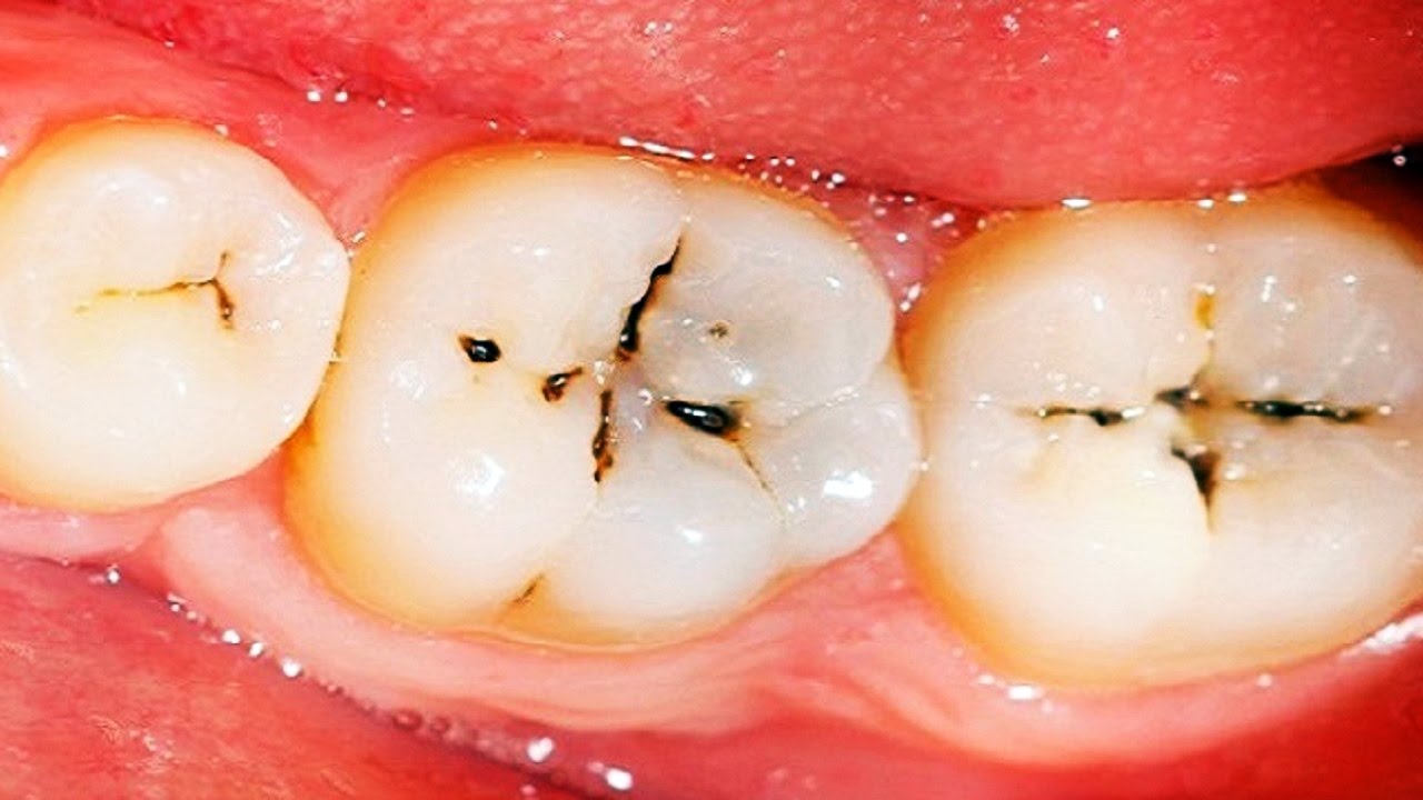
Cavities often appear smaller than the decay underneath
Surface holes can hide deep internal damage. X-rays reveal the truth. What looks minor may span the tooth. Restoration requires depth. Fillings grow larger than expected. Infections start invisibly. Visual inspection isn’t always enough.
Bacteria exploit dentinal tubules to reach the pulp chamber
These microscopic tunnels guide invaders. Pulp houses nerves and vessels. Once reached, pain begins. Inflammation rises. Pressure builds inside. Pulsing follows. The tooth becomes temperature-sensitive and pressure-reactive. This stage pushes many to the dentist.
Pulp inflammation doesn’t always mean immediate death of the tissue
Some pulps survive mild inflammation. Others deteriorate quickly. It depends on immune response and pressure. Swelling has no exit. Blood vessels collapse inside the chamber. Necrosis begins. Tissue darkens. Infection follows. Pain may stop as nerves die—but damage grows.
Infection can escape the tooth and reach the surrounding bone
Once the pulp dies, bacteria exit through the root tip. Abscesses form. Swelling spreads. Gums redden. Jaws ache. Cheeks may swell. Lymph nodes react. The infection leaves the tooth’s structure. It travels under soft tissue layers.
Swelling under the jaw may signal deep dental abscess spread
Facial asymmetry suggests urgency. Breathing or swallowing difficulty requires attention. Antibiotics become necessary. Drainage may follow. Hospitalization isn’t rare in neglected infections. The mouth becomes a gateway for systemic issues.
Pain may disappear briefly before worsening with deeper infection
As nerves die, pain fades. This lull misleads. Pressure builds. Pus accumulates. Symptoms return stronger. Throbbing escalates. The illusion of improvement hides deepening infection. Waiting at this point allows wider spread.
Bone loss begins when inflammation reaches the periodontal ligament
The ligament surrounds roots. Inflammation here weakens bone. Support structure shrinks. Teeth loosen. Chewing hurts. X-rays show gaps between bone and root. Gum pockets deepen. The tooth destabilizes without trauma.
Chronic infection can drain silently through gum fistulas
Small bumps may form near roots. Pus escapes. Pain lessens temporarily. But the cycle continues. Fistulas act like vents. They don’t solve the issue. Internal infection persists. These signs require endodontic care—not monitoring.
Decayed teeth may not smell but harbor active pathogens
Many think odor equals infection. But early decay may carry none. Bacteria thrive below surfaces. They colonize tubules. Brushing masks presence. Only close inspection reveals depth. Fresh breath doesn’t rule out problems.
Cavities between teeth often develop without visible signs
Interproximal decay hides well. It spreads under contact points. Flossing pain may be the first sign. X-rays catch it late. Clean outer enamel misleads. These cavities grow sideways until the wall collapses.
Root surfaces decay faster due to thinner protective layers
Exposed roots lack enamel. They rely on cementum—a softer barrier. Aging, gum disease, and brushing recession expose roots. Once visible, they decay quickly. Acid wears cementum faster. Cold air triggers pain. Root cavities complicate fillings.
Secondary decay can form under old dental restorations
Marginal breakdown lets bacteria re-enter. Fillings don’t last forever. Edges lift. Gaps form. Decay returns silently. Regular exams reveal soft spots. Retreatment becomes necessary. New decay under fillings causes confusion.
Recurrent cavities spread faster due to weakened surrounding tissue
Previous fillings reduce tooth strength. New decay nearby progresses rapidly. Margins fail first. Restoration integrity fades. Small weaknesses become large restorations. Repeat treatment becomes deeper, wider, less conservative.
Deep decay near the pulp may require indirect pulp capping
If decay nears but doesn’t enter pulp, dentists try to preserve tissue. They place medicine below filling material. Healing is possible. But follow-up matters. Pain later may signal failure. Close margins require monitoring.
Once the root is infected, root canal therapy becomes necessary
Endodontic treatment removes infected pulp. Tools clean canals. Irrigation disinfects. The tooth remains, but nerves go. Pain resolves. Structure stays. It’s conservative compared to extraction. But success depends on infection limits.
Root canals don’t guarantee lifetime tooth survival
Post-treatment fracture risk increases. Teeth become brittle without blood supply. Crowns reduce breakage. Monitoring matters. Incomplete sealing invites reinfection. A well-treated tooth lasts years—but not always forever.
Untreated decay in children can affect permanent tooth development
Baby tooth infection reaches bone. The adult tooth forms underneath. Inflammation alters enamel. Spots appear later. Sometimes eruption delays. Primary decay isn’t harmless. It transfers effects downward.
Small children may not express early decay pain clearly
Night wakings. Avoided foods. Drooling. These signs suggest dental discomfort. They precede speech complaints. Visual checks miss hidden decay. Fluoride helps—but timing matters. Intervention before speech clarity matters most.
Fluoride prevents demineralization but doesn’t reverse cavity formation
It hardens enamel. It strengthens weak spots. But it doesn’t rebuild missing structure. Cavities still need fillings. Fluoride reduces new damage—not heals holes.
Remineralization efforts only help in earliest decay phases
White spot lesions can heal with minerals. Once cavitation starts, restoration becomes necessary. Early treatment catches reversibility. Delayed care closes that option. Prevention stays time-sensitive.